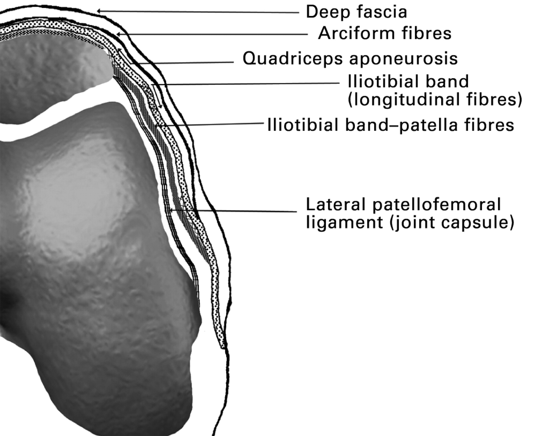
Medial Retinaculum Anatomy

The medial retinaculum is a thick, fibrous band that forms the palmar boundary of the carpal tunnel. It is located on the volar aspect of the wrist, extending from the pisiform bone to the hook of the hamate bone. The medial retinaculum is attached to the transverse carpal ligament proximally and to the palmar carpal ligament distally. It is innervated by the median nerve.
The medial retinaculum plays an important role in maintaining the integrity of the carpal tunnel. It prevents the tendons of the flexor muscles from bowing out of the carpal tunnel, which would compress the median nerve. The medial retinaculum also helps to protect the median nerve from injury.
Attachments
The medial retinaculum has three main attachments:
- Proximally, it is attached to the transverse carpal ligament.
- Distally, it is attached to the palmar carpal ligament.
- Laterally, it is attached to the hook of the hamate bone.
Innervation, Medial retinaculum
The medial retinaculum is innervated by the median nerve. The median nerve provides sensory innervation to the palmar aspect of the hand, including the thumb, index finger, middle finger, and ring finger. The median nerve also provides motor innervation to the muscles of the thenar eminence, which are responsible for thumb movement.
Medial Retinaculum Function

The medial retinaculum plays a crucial role in facilitating hand movements, preventing bowstringing of the flexor tendons, and contributing to the overall stability of the wrist joint.
The medial retinaculum acts as a pulley, holding the flexor tendons close to the wrist bones. This allows the tendons to exert a more efficient force on the fingers, enabling smooth and controlled hand movements. Without the medial retinaculum, the tendons would bowstring, reducing their mechanical advantage and impairing hand function.
Preventing Bowstringing of Flexor Tendons
The medial retinaculum prevents bowstringing of the flexor tendons by creating a tunnel-like structure that keeps the tendons in place. This prevents them from bulging out when the wrist is flexed, which could impair their function and lead to pain.
Contribution to Overall Wrist Joint Stability
The medial retinaculum also contributes to the overall stability of the wrist joint. It acts as a stabilizing band, preventing excessive lateral movement of the carpal bones and providing support to the wrist ligaments.
Medial Retinaculum Pathology
The medial retinaculum can be affected by various conditions, including carpal tunnel syndrome and De Quervain’s tenosynovitis. These conditions can cause pain, numbness, and weakness in the wrist and hand.
Carpal tunnel syndrome is the most common condition that affects the medial retinaculum. It occurs when the median nerve, which runs through the carpal tunnel, is compressed. The carpal tunnel is a narrow passageway in the wrist that is formed by the bones of the wrist and the medial retinaculum.
Carpal Tunnel Syndrome
- Causes: Carpal tunnel syndrome can be caused by a variety of factors, including repetitive hand movements, obesity, pregnancy, and certain medical conditions, such as diabetes and rheumatoid arthritis.
- Symptoms: The most common symptoms of carpal tunnel syndrome include pain, numbness, and tingling in the thumb, index, middle, and ring fingers. The pain may be worse at night or when the hands are used for prolonged periods of time.
- Treatment: Treatment for carpal tunnel syndrome may include conservative measures, such as splinting, corticosteroid injections, and physical therapy. In severe cases, surgery may be necessary to release the pressure on the median nerve.
De Quervain’s Tenosynovitis
De Quervain’s tenosynovitis is another condition that can affect the medial retinaculum. It occurs when the tendons that control the thumb movement become inflamed. The tendons run through a narrow passageway in the wrist called the first dorsal compartment, which is formed by the bones of the wrist and the medial retinaculum.
- Causes: De Quervain’s tenosynovitis is most commonly caused by overuse of the thumb, such as in activities that involve repetitive gripping or pinching motions.
- Symptoms: The most common symptom of De Quervain’s tenosynovitis is pain at the base of the thumb. The pain may be worse when the thumb is used for gripping or pinching.
- Treatment: Treatment for De Quervain’s tenosynovitis may include conservative measures, such as splinting, corticosteroid injections, and physical therapy. In severe cases, surgery may be necessary to release the pressure on the tendons.
The medial retinaculum, a connective tissue in the wrist, ensures smooth hand movements. Like Theresa Randle’s captivating performance in “Bad Boys 4” theresa randle bad boys 4 , the medial retinaculum plays a crucial role in our everyday actions. Just as Theresa’s character adds depth to the film, this ligament stabilizes the wrist joint, allowing us to effortlessly reach, grasp, and move.
The medial retinaculum, a band of connective tissue that holds tendons in place, plays a crucial role in wrist stability. Like the strategic guidance provided by the owner of the Dallas Mavericks , it ensures smooth and controlled movement. Understanding its function empowers us to appreciate the delicate balance that underlies our physical capabilities, much like the interplay between leadership and team performance on the court.
The medial retinaculum, a ligament in the wrist, provides stability and support to the carpal bones. It’s fascinating how its role in maintaining wrist function parallels the comedic prowess of George Lopez, whose stand-up routines in Eagle Mountain sent audiences into fits of laughter.
Just as the medial retinaculum ensures smooth wrist movements, Lopez’s wit and timing orchestrate a symphony of chuckles.
Like a medial retinaculum, a delicate yet crucial band that stabilizes the tendons of the hand, the connection between jennifer hudson and common has been a stabilizing force in both their personal and professional lives. Just as the medial retinaculum enables seamless movement, their relationship has provided support and inspiration for their creative journeys, allowing them to explore new dimensions of their artistry.
The medial retinaculum, a connective tissue structure, plays a pivotal role in stabilizing the wrist. Interestingly, its intricate network of fibers has been compared to the masterful play of basketball legend Bill Walton , who seamlessly navigated the court with precision and agility.
Just as Walton’s footwork anchored his dominance, the medial retinaculum provides a secure foundation for the intricate movements of the wrist.